The Key to Early Diagnosis in Heart Diseases: Cardiac CT and MRI
Assoc. Prof. Dr. Vefa Çakmak, a faculty member of the Department of Radiology at Pamukkale University (PAU) Hospitals, explained the advantages of Cardiac CT (Computed Tomography) and Cardiac MRI (Magnetic Resonance Imaging), which have become increasingly important in recent years for the diagnosis of heart diseases.
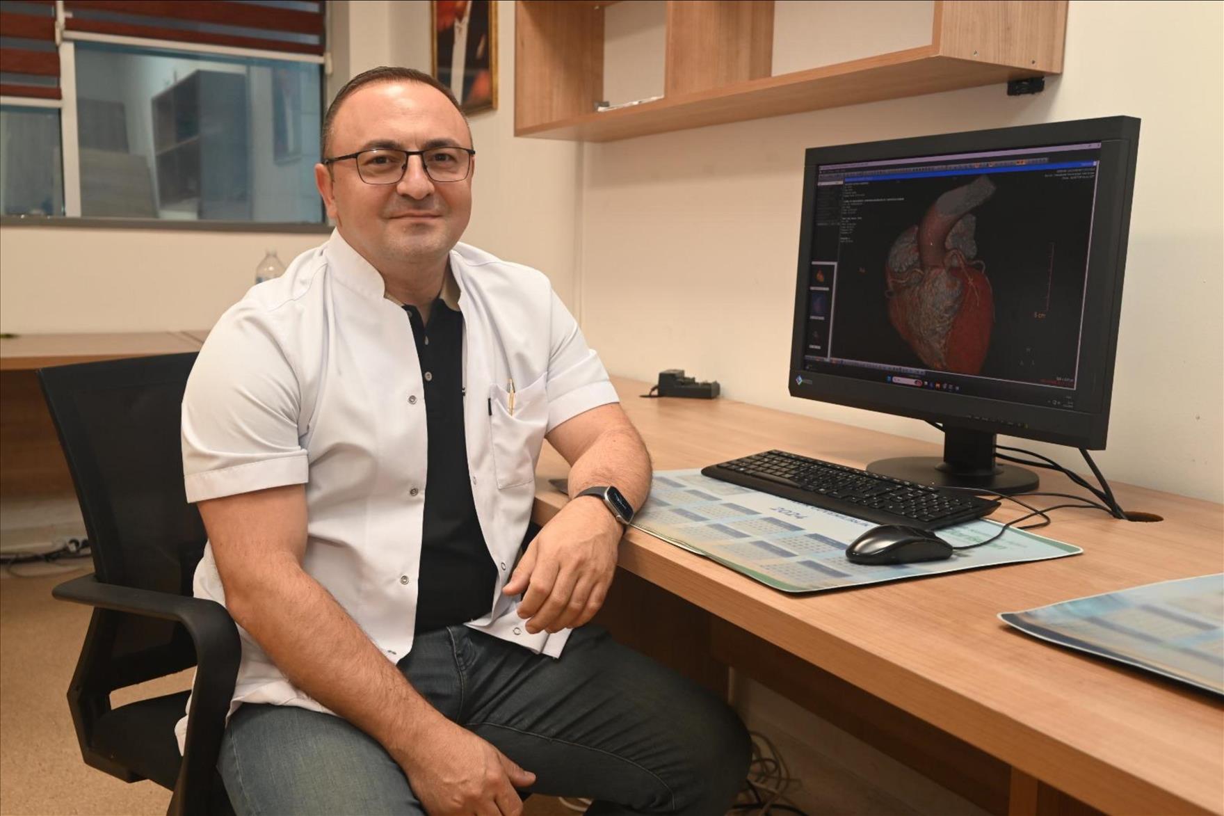
Assoc. Prof. Dr. Vefa Çakmak stated: “Computed tomography (CT), which became more common in our lives during the pandemic, continues to be used as a vital imaging method in diagnosis and treatment processes today. This radiation-based technology allows detailed examination of many organs and provides rapid diagnosis in emergencies, playing a life-saving role. It is particularly significant in cancer diagnosis and follow-up, traffic accidents, brain hemorrhages, fractures, and preoperative assessment for major surgeries. CT enables detailed imaging of the heart’s blood vessels. This method, commonly referred to as a ‘virtual angiography,’ allows early detection of coronary artery blockages that may require intervention, thus protecting patients from unnecessary traditional angiography procedures. At the same time, heart walls, thrombi or tumors inside the heart, and structures that may cause arrhythmia can also be evaluated. Additionally, low-dose lung imaging can be performed during the scans, allowing detection of structural lung diseases, lung cancers, and thoracic skeletal issues.
Before the scan, blood tests such as creatinine and GFR are performed to protect kidney function. The patient’s medical history and drug allergies are also reviewed. To obtain high-quality images, the heart rate may need to be reduced to around 70 beats per minute; in such cases, heart rate-lowering medications may be used. During the procedure, the patient is monitored, metallic items on the chest are removed, and a breath-hold of approximately 10–15 seconds is requested. Cardiac CT scanning takes 4–10 seconds, and the images are usually evaluated and reported on the same day.”
Assoc. Prof. Dr. Vefa Çakmak: “The lung condition of smokers can be clearly evaluated.”
Highlighting the method’s ability to reveal the presence or absence of major conditions such as heart disease, vascular blockage, lung disease, or cancer, Assoc. Prof. Dr. Çakmak continued: “Especially in smokers, the impact on the lungs can be clearly seen. This answers the frequently asked question of smokers: ‘Could I have cancer?’ The procedure has no harmful effects, and the technology used complies with international standards.
Another important method in diagnosing heart diseases, Cardiac MRI, is used in cases where angiography cannot provide a diagnosis. It is preferred for the detection and follow-up of structural heart wall diseases, heart valve problems, iron accumulation, and other specific conditions. This method does not involve radiation. During the approximately 25–30 minute scan, patients remove metallic items, are monitored, and follow breath-holding instructions. The scan is performed according to the request of the relevant physician, and results are usually delivered to the patient within one business day. Both CT and Cardiac MRI imaging are conducted by expert teams, and the data obtained provide highly valuable information for diagnosis. These technologies allow many diseases to be detected early, prevent unnecessary interventions, and ensure the highest level of patient safety.”